Memories of Professor Keiichi Tanaka
Tottori University Emeritus Professor Keiichi Tanaka, known around the world as an authority on scanning electron microscopy (SEM)—passed away in October 2019 at the age of 93.
In 1966, Professor Keiichi Tanaka had an opportunity to use the first SEM system sold in Japan, which he used to capture SEM images of his research focus at the time: crystalline structures in the eye. Electron microscopy in those days was considered unsuitable for biological samples with substantial water content, but Professor Tanaka had the ingenious idea of infusing glycerol into samples, and in this way successfully acquired the first SEM images of biological samples in Japan. Thereafter, Professor Tanaka shifted the focus of his research to applying SEM to biological samples and increasing the associated magnification; in 1985, while serving as the Dean of Tottori University’s Faculty of Medicine, Professor Tanaka worked with Hitachi to develop—after many years of dedicated effort—an SEM system with 800,000×magnification, the highest of any instrument in the world at that time. This microscope was used to capture clear images of the AIDS virus, which were distributed around the world and became a focus of intense interest.
Thus, it was Keiichi Tanaka who opened the door to the development of SEM for life-science applications. To commemorate this accomplishment, we organized a special online conversation with Tottori University’s Sumire Inaga, a trusted protégé of Professor Tanaka, and Daisuke Koga of Asahikawa Medical University, who has continued the development of sample-fabrication methods pioneered by Professor Tanaka. In this first part of our conversation, titled “Memories of Professor Keiichi Tanaka,” our panelists shared anecdotes from their work with Professor Tanaka and discussed their current research.
Note: This program is divided into Part One and Part Two.
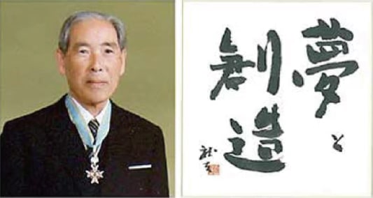
Professor Keiichi Tanaka
Former Dean of the Tottori University
Faculty of Medicine
Recipient, Order of the Sacred Treasure
and Yonago City Citizen’s Honor Award
(1997)
INAGA: I expect you’re already familiar with Professor Tanaka’s career, but let me start by just quickly reviewing it. He graduated from Yonago Medical University, the predecessor of today’s Tottori University Faculty of Medicine, and, after a one-year internship, became a research associate in the Department of Anatomy. From that moment until his retirement in 1991, he devoted himself to research and teaching in the Department of Anatomy, and it was while serving as Dean of the Faculty of Medicine that he developed the UHS-T1 ultra-high-resolution scanning electron microscope. I regret to say that at that time I was out on parental leave, and thus cannot personally recount the step-by-step process leading up to the development of this instrument, but Professor Tanaka described it in detail in his book “The challenge of the ultramicroscopic world: Seeing living organisms at 800,000×magnification.”
Technically, my affiliation was not with Professor Tanaka’s group—the Second Anatomy Group—but rather with the First Anatomy Group led by Professor Akihiro Iino, a former member of Professor Tanaka’s group. Nonetheless, the members of our two groups treated each other like family, and we pursued our research with a spirit of friendly collaboration.
The focus of my research was elucidating the higher-order structure of chromosomes, and, when I returned to work one year after the ultra-high-resolution SEM was completed, Professor Tanaka asked me to help verify the performance of the new instrument by observing the double-helix structure of DNA. Even though Professor Tanaka graciously volunteered his own time as an operator to help me tackle this challenge, at first things didn’t go very well. But, after about a year and a half of repeated trial-and-error experimentation, we finally captured clear images of the double-helix structure—I still remember to this day how thrilled I was to see those images.
One event that always left a strong impression on me was Professor Tanaka’s decision to step down as Dean of the Faculty after just a single two year term. It was traditional for Deans to serve two terms, but Professor Tanaka simply said, “I’m eager to get back to my research,” and resigned as Dean—no doubt to the regret of many of his peers. I remember clearly what he said at the time, “I want to pilot fighter planes, not passenger jets!” That was Professor Tanaka’s unwavering commitment to pursuing cutting-edge research. My own feeling was something like “Just sitting in the corner of that fighter plane is more than enough for me—let me join you on your journey!” And I gave everything I had to accompanying Professor Tanaka on his mission.
KOGA: That sounds like Professor Tanaka to me. He must have been greatly admired by everyone who worked with him.
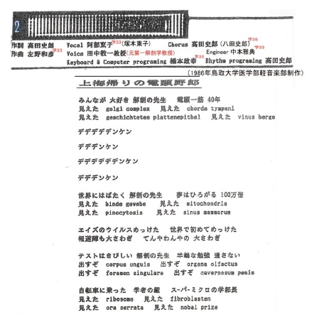
The lyrics to a song about Professor Tanaka written by Tottori University students.
One gets a sense of the affinity with which the students viewed their famous mentor.
INAGA: Oh, yes. When the ultra-high-resolution SEM was completed, some students in the University’s Pop Music club composed and wrote lyrics for a song celebrating the achievement, which they gave Professor Tanaka as a gift. It was a fun song called “The electron-microscope guy is back from Shanghai,” and it had lyrics like “He’s the anatomy professor that everybody loves” and “The very image of the scholar on a bike, whoa! He’s the Dean of the super-micro!” In contrast to most people at that time, who commuted by car, Professor Tanaka rode back and forth to campus on his bicycle, and at lunchtime every day he would ride home to enjoy his wife’s home cooking. Apparently, students would frequently see him riding his bike and found the sight adorable.
KOGA: I can easily picture that! I didn’t start down the research path until after Professor Tanaka had retired, so I regret that, unlike Professor Inaga, I can’t offer personal recollections of his years at Tottori, but I definitely remember that his paper on the osmium-maceration method was a major turning point in my career. When I first saw the cross-sectional images in that paper on cells captured by osmium maceration, I remember being totally overwhelmed, and thinking, “Wow, SEM is really capable of visualizing a whole new world!” Whereas SEM had previously been limited to imaging the exterior of cells, this new method enabled the world’s first direct visualization of structures inside cells, including mitochondria and the endoplasmic reticulum. I decided right away that I wanted to replicate this fantastic technique on my own.
However, although I tried to follow the method as described in the paper, things didn’t go very well. I spent day after day searching by trial and error on my own to find the ideal parameter settings that would lead to successful images—or to discover the key trick that wasn’t mentioned in the paper. Eventually, during my years as a graduate student, I finally figured things out, and images of cells that I acquired via osmium maceration were featured on the cover of a certain scientific journal four times in a single year. This journal also carried a column by Professor Tanaka, and he must have graciously taken the time to look at my images, because after the first one was published I received a letter from him.
In the letter, he had written, “I saw your image. You did an excellent job!” I remember reading that compliment and feeling truly overjoyed. I immediately wrote back, and thereafter Professor Tanaka and I continued exchanging letters right up until he passed away. Back when I was a graduate student and my research trajectory was still very uncertain, he went out of his way to express warmth and take me under his wing. And, after that, whenever I was having trouble in my research and asked him for advice, he would always give me helpful suggestions—he really provided spiritual support in that way.
I’ll also never forget when I visited his home in Tottori and enjoyed Mrs. Tanaka’s delicious home cooking. To me, the opportunity to interact with someone like Professor Tanaka—someone you could really respect, both as a researcher and as a human being—was an irreplaceable treasure.
INAGA: I think it’s very impressive that you were able to master the osmium-maceration method on your own!
KOGA: Thank you. You might call it stubbornness on my part, but asking Professor Tanaka to teach me about his method always seemed wrong to me. To me, the most important thing was to succeed in fabricating clean samples entirely on my own—and then to have Professor Tanaka praise my work. That was the goal on which I focused all my effort.
Nowadays, I myself get asked all kinds of questions, but I tend not to like it when students ask me to explain everything from the very beginning. It’s important for students to get their own hands self-studying around with things first! I’m hoping to pass this point of view on to the next generation of researchers as well.
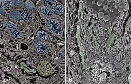
Example of an osmium-maceration SEM image
THE HITACHI SCIENTIFIC INSTRUMENT NEWS Vol.7 (2016)
Serial-section scanning electron microscopy
https://www.hitachi-hightech.com/global/en/sinews/si_report/07054/
INAGA: Professor Tanaka himself was not the type of person to say this or that about imaging methods or other techniques. But he was extremely strict when it came to getting results—he was always saying “no matter what, be prepared at all times to take beautiful pictures.” I really think beauty was the most important criterion in his mind. The attitude that “as long as the image is true, that’s all that counts” is common in electron microscopy, but Professor Tanaka really cared about the composition of the image—“if the picture isn’t beautiful, it won’t convince anybody,” he used to say. I used to show him images I had captured, and he would say “Hmmm, your image composition is a little lacking—why don’t you go to an art museum and look at some paintings to see how it’s done?” And I would go back and recapture the image, or if I couldn’t recapture the image I would try to do something clever with the trimming or something like that, and eventually I came to appreciate the importance of beautiful images on my own terms.
The English poet John Keats famously wrote, “Beauty is truth, truth beauty.” Professor Tanaka took these words to heart, and they lay at the root of all his value judgments.
I mentioned before that it took about a year and a half to image the double-helix structure of DNA. The fact that Professor Tanaka was even willing to wait that long testifies again to his unwavering commitment to beauty—and to the depth of his resolve. Nowadays everything is always expected to be delivered in speedy fashion, but I think it’s time we took a step back and remembered Professor Tanaka’s strict attitude toward high-quality results—and the patience he showed in being willing to wait for them.
KOGA: With regard to the idea of beauty, I feel the same way—if you’re going to capture images you should insist on capturing beautiful ones. Obviously, this is particularly essential if you’re a morphologist, but I think many of the pioneers of electron microscopy, not just Professor Tanaka, were sticklers for beauty as well. For example, no matter how important a sample may be, if the sample has any dirt on it I simply won’t take a picture of it, no matter what. Instead, if possible I’ll make a new sample all over again from scratch. But, in practice, I think relatively few people insist on beauty. As Professor Inaga says, these days all the focus is on getting results quickly. This may be inevitable in a results-driven world, but I do think that among electron microscopists the philosophical aspect of our work—the question of what do I really want to show?—is getting short shrift.
To improve this situation, I think it would be ideal if there were more opportunities to learn things like electron-microscope imaging techniques. Even if that proves too difficult, I myself plan to continue insisting on taking beautiful images—and in this way to communicate the beauty of our field to the rest of the world.
INAGA: I retired from the University in March 2018, and since then I’ve been working as a project researcher to continue my collaborative research with Hitachi High-Tech. The main focus of my research is establishing a diagnostic method for renal biopsies using low-vacuum tabletop SEM systems.
I started analyzing pathological tissue in the kidney i n 2008, but i n fact for some time before that I had been working on low-vacuum SEM observations of pathological tissue. Low-vacuum SEM allows observation of samples with high water content such as living tissue, as well as electrically insulating samples, without complicated preprocessing, and the simplicity of the procedure got me wondering why we couldn’t use it for biopsies. However, I’m not a clinician, so I wasn’t sure which kinds of tissue would be the most desirable targets for something like this. That was when a colleague from Hitachi High-Tech who worked on transmission electron microscope(TEM) said, “Why don’t you look at kidney tissue? Because there’s already an established TEM-based diagnostic method for that.” So, I shifted my focus to renal biopsies.
At the time, I was just a research associate and didn’t have much research funding, so I borrowed lab space and prototype instruments from Hitachi High-Tech and collected slides from pathologists and kidney specialists, which I was able to organize into several publications. This work was not immediately recognized by clinicians, but my colleague from Hitachi High-Tech and I worked together to give a series of solid presentations at microscopy conferences and anatomy conferences, and eventually my work started to attract some interest.
One of the first people to pay attention was Emeritus Professor Nobuaki Yamanaka of Nippon Medical School, a well-known authority on kidney pathology, who graciously agreed to chair the Renal Biopsy LVSEM Research Group* that we established in 2017. Since then, a number of other professors have recognized the promise of our work, and we’re currently moving forward toward establishing a diagnostic method with clinical applications. Although it’s taken 10 years, I feel blessed that my research has gotten to see the light of day and seems increasingly likely to be of value to society.
Another focus of my research, also in collaboration with Hitachi High-Tech, is the use of focused Ion beam (FIB) systems to study the higher order structure of chromosomes. As I mentioned previously, this is something I’ve been working on for more than 40 years, and the reality is that, although the decoding of the DNA base sequences has progressed during that time, the detailed structure of chromosomes is still not clearly understood. Shedding light on this subject is one of the goals of my research.
Examination of the light microscopic slide of renal biopsy specimens by utilizing Low- vacuum scanning electron microscope
https://www.hitachi-hightech.com/global/en/sinews/si_report/090202/
INAGA: Absolutely. There will be several benefits, of which perhaps the most important will be a reduced burden on patients. In general, observation and diagnosis of living tissue—not just kidney tissue—requires collecting tissue samples and preparing paraffin sections for optical microscopy. For kidney tissue, diagnosis via TEM is also possible, but this requires collecting new samples separate from the samples used to prepare paraffin sections. In contrast, diagnosis via low-vacuum SEM can be done with standard paraffin sections prepared as usual. This is the greatest advantage of the technique.
Also, TEM-based electron-microscope diagnosis requires specialized knowledge and expertise to prepare samples and make diagnoses, which usually entails outsourcing to a specialty testing company—which is expensive and time-consuming. Low vacuum SEM systems are sufficiently inexpensive and easy to use, so they can be installed in any medical institution and used for routine pathology screening.
There has also been progress in methods for staining samples. In the early days, a signal amplifying agent called platinum blue—developed by Professor Tanaka—was used, but since then we’ve learned that observations are also possible using the same periodic acid-methenamine-silver(PAM) staining process commonly used for paraffin sections. This means that samples prepared for observation via conventional approaches can be observed as-is via SEM, yielding more fine-grained data than are available via optical microscopy and promising earlier diagnosis of pathologies. In particular, with the frequency of kidney transplants on the rise in recent years, this method will allow earlier detection of—and response to—transplant rejections, i.e., symptoms of transplanted kidneys being rejected due to immune response. This will also be a major benefit.
KOGA: There are a number of 3D imaging techniques for analyzing the ultra-structure and morphology of living organisms, but the ones I’m focusing on presently are osmium maceration and serial-section SEM. Serial-section SEM is a brand-new technique in which a few hundred to a few thousand ultra-thin sections are sliced from a specimen embedded in resin and these slices are observed in sequence via SEM, with any regions of particular interest analyzed via 3D reconstruction. I believe our group was the first in Japan to successfully establish this method.
Osmium maceration is a technique in which the so-called freeze-fracture method is used to prepare clean slices of cells, the soluble proteins are then removed from the cell cross section, thereby selectively retaining only the cell membrane. This is a very attractive imaging technique, and it is a powerful tool for ultra-high-resolution 3D imaging of local features when observing cell cross sections—but it is not ideally suited to capturing full body images of organelles that extend throughout the cell interior. I am particularly interested in the organelles known as Golgi apparatuses, which are much larger than mitochondria and sometimes even nuclei, and which cannot be fully observed just by looking at cross-sectional images. Professor Tanaka used osmium maceration to observe Golgi apparatuses, but this approach is best suited to observations of detailed structure in the Golgi cisternae that layer together to comprise the Golgi apparatus. In contrast, I felt strongly that I wanted to see full-body images of Golgi apparatuses, which is difficult to do via osmium maceration—and which led me to try serial-section SEM.
When you think of a Golgi apparatus, the image that comes to mind is probably the common textbook diagram of bag-like Golgi cisternae piled atop one another. However, as was demonstrated in an article in SI NEWS*, the actual 3D structures of Golgi apparatuses are complicated—and nothing like the textbook diagram. Thus far we have shown that the shapes of Golgi apparatuses differ significantly depending on cell type and functional state; going forward we are working to understand the basic shapes of Golgi apparatuses in a wide variety of cells, and we are hopeful that this research will prove useful for understanding pathologies characterized by morphological changes in these organelles.
https://www.hitachi-hightech.com/global/en/sinews/si_report/07054/
KOGA: Performing serial-section SEM observations in practice requires developing a stable slicing technique for preparing ultra-thin sections of samples. When choosing observation subjects, it’s also important to keep in mind that the thickness of the ultra-thin sections is closely related to the spatial resolution in the z-direction.
Aside from serial-section SEM, other 3D imaging techniques include FIB-SEM, in which an FIB system is used to shave down a sample surface while observing it. This approach can cut resin-embedded sample blocks with a pitch of a few nanometers, yielding high z-axis resolution. On the other hand, because this method is destructive, repeating previously-captured images is impossible. The same is true for another method: serial block-face (SBF)-SEM, in which a diamond knife installed inside the SEM specimen chamber is used to cut sample cross sections as they are observed. Serial-section SEM has the advantage of enabling samples to be preserved semi-permanently, allowing images to be captured and re-captured any number of times.
Other advantages of serial-section SEM include the ability to analyze wide sample regions by enlarging the size of the section block and the fact that observations can be made with any high resolution SEM system, without the need for any specialized equipment. Of course, every method has its advantages and disadvantages, and the best strategy is to choose the optimal method based on the subject and goals of your observation.
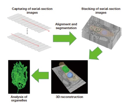
Procedure for 3D reconstruction of serial-section images
THE HITACHI SCIENTIFIC INSTRUMENT NEWS Vol.7 (2016)
Serial-section scanning electron microscopy
https://www.hitachi-hightech.com/global/en/sinews/si_report/07054/
KOGA: Yes, it seems many people find that to be a major hurdle to overcome. In our case, it took almost 2 years to develop our technique, but we were absolutely committed to viewing full-body images of Golgi apparatuses no matter what, so we worked with Assistant Professor Satoshi Kusumi of Kagoshima University for as long as it took to achieve our objective. But isn’t that what research is all about? You are constrained by various limitations, but you focus on what you want to learn and use all your knowledge and energy to get there. For us, as long there’s some object—some structure—that we want to observe via SEM, we will spare no effort to make that observation.
For those of us who are following in Professor Tanaka’s footsteps, the crucial mission is to develop the SEM system that he built with Hitachi High-Tech as an academic tool. Of course, Professor Inaga didn’t stop there, but pressed onward to extend the power of electron microscopy into the domain of clinical testing, which I think is absolutely fantastic.
INAGA: Yes, even Professor Tanaka was surprised and delighted to learn about clinical applications of low-vacuum SEM. Of course, Professor Tanaka focused on the potential of low-vacuum SEM from an early stage—he started researching applications after he retired from the University, some 26 or 27 years ago. He was convinced that the advantages of the low-vacuum SEM, which can be installed even at home and used to observe biological samples with high water content, had the potential to open up brand new worlds of discovery. In April 2000, he opened the Tanaka SEM Laboratory in his home, and for many years he was a devoted user of the S-2460 Natural SEM.
When Professor Tanaka first became interested in low-vacuum SEM, he was ahead of his time, and his work didn’t attract much interest. But, more recently—and thanks in large part to Hitachi High-Tech’s work developing new systems—low-vacuum SEM has become a ubiquitous tool. I’m convinced that the availability of tools that were easy to use played a key role in expanding the world of potential applications to include medical diagnoses and other new domains.
Continuing the Mission of Professor Keiichi Tanaka
A conversation with Sumire Inaga of Tottori University and Daisuke Koga of Asahikawa Medical University to commemorate the accomplishments of Professor Keiichi Tanaka, an international authority on SEM who passed away in October 2019.
In this second part of our conversation, titled “Continuing the Mission of Professor Keiichi Tanaka,” we asked our panelists to describe their efforts to advance Professor Tanaka’s lifelong mission to expand the possibilities of electron microscopy—and to discuss future prospects for their own research.
(Panelists)
Sumire INAGA
Project Researcher
Department of Anatomy
Tottori University Faculty of Medicine
Daisuke KOGA
Associate Professor
Department of Microscopic Anatomy and Cell Biology
School of Medicine Medical Course Basic Medicine Anatomy
Asahikawa Medical University
INAGA: This is something that arose out of the blue just as I was retiring from the university in 2018. It all started when Professor Tanaka’s advanced age started to make it difficult for him to continue his research, and he decided to donate his cherished S-2460 Natural SEM system—which he had installed at his home and used with great pleasure for many years—to Yonago City. It seems that initially there was some progress toward making this happen, but then things ground to a halt due to various obstacles and a change of mayors, and eventually Professor Tanaka asked me what would be best to do. At that point I asked one of my former classmates to step in and help out, and we wound up contacting the city’s Superintendent of Education to rejuvenate the project.
As it turned out, Professor Tanaka was not the only giant of microscopy to hail from Yonago City: the late Professor Eizi Sugata, a TEM pioneer who was Dean of the Osaka University School of Engineering, was from Yonago as well. Thus Yonago can count the preeminent researchers in both SEM and TEM among its native sons—an honor that links the city inextricably to the art and science of microscopy, but one which remains almost entirely unknown! So we used this opportunity to help spread the word: we created an exhibition showcasing the microscope that Professor Tanaka donated and other instruments, both to celebrate and tell the world about the achievements of Yonago’s two illustrious microscopists and to establish a meaningful environment for microscopy education—we even proposed holding a fund-raising drive to allow us to purchase a new microscope system. And thus, with the enthusiastic support of Yonago’s mayor, we launched our project to install a tabletop low-vacuum SEM system—both for exhibition purposes and for actual research—in a corner of the Yonago City Children’s Center.
As for my part in all this, I had been planning to wait until I retired and had more free time before getting involved, but the Dean of the Faculty of Medicine said “No, the alumni and the Medical Association are willing to help out, so you should really take care of it before you retire.” So I started working on it in December 2017. As it turned out, we received fewer donations than we had expected, and we didn’t achieve our funding goal of 5 million yen until after the opening ceremony on March 26, 2018—a rather precarious situation! Happily, though, Professor Tanaka—who had been hospitalized for more than 2 months at that point—was able to attend the ceremony in a wheelchair, and we were thrilled to be able to celebrate the occasion with him.
Our actual Miniscope® system is installed in one corner of the main entrance hall of the Children’s Center; we call it the Corner for Exploration of Microscopic Worlds and the installation is designed to make it easy for anybody—both adults and children—to observe microscopic phenomena. The system can only be used when a supervisor is present—we have a total of three supervisors, including myself—and we do get many requests from people asking us to show them a specimen. During summer vacation we allow users to sign up for one-hour slots during which the instrument is theirs to use for their independent research; there are a total of about 40 such slots, and they always get fully booked almost immediately. The total number of electron-microscopy images captured to date is already around 9,000.
KOGA: I’ve visited this facility too—I was invited to speak as part of a special lecture series commemorating its dedication, and I remember being astonished by the unusual laboratory environment. It was the first time I had ever seen an electron microscope installed next to a pingpong table! But the notion of a facility offering both children and adults the opportunity to play around with electron microscopy is truly revolutionary, and I doubt there’s anything else like it in the world.
INAGA: Even though the system is installed in an environment with a lot of dust and a lot of vibrational noise—and with children running a round everywhere—to date we’ve had no operational problems with the instrument itself; all that dust seems to have no impact on the interior of the microscope! It’s a real testament to the solid construction and robustness that Hitachi High-Tech designed into the Miniscope® system.
*Promotional website for Yonago: the electron-microscope town:
https://denkenyonago.com/
(in Japanese)
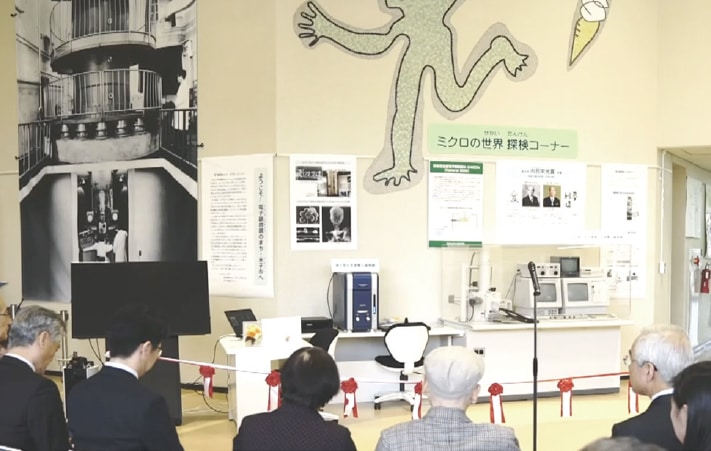
The Corner for Exploration of Microscopic Worlds at the Yonago City Children’s Center.
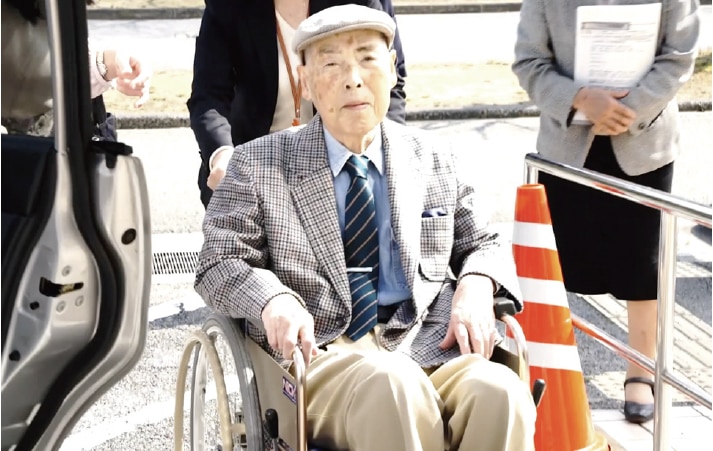
Professor Tanaka Keiichi in good spirits at the opening ceremony.
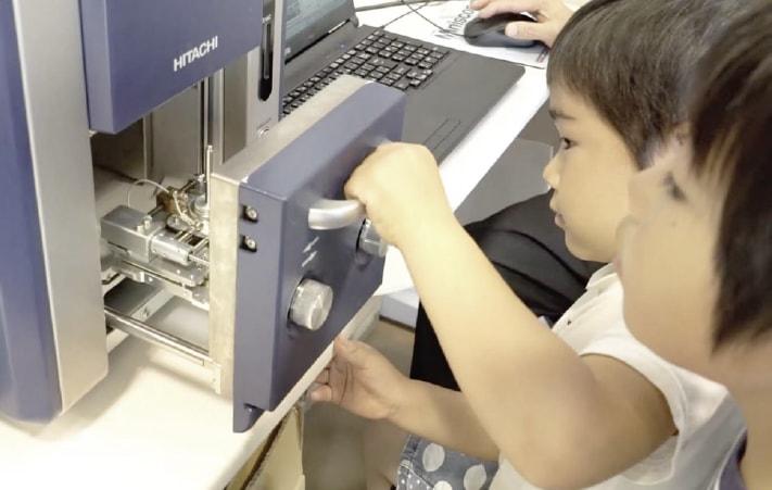
Children have the opportunity to control the electron-microscope system with their own hands.
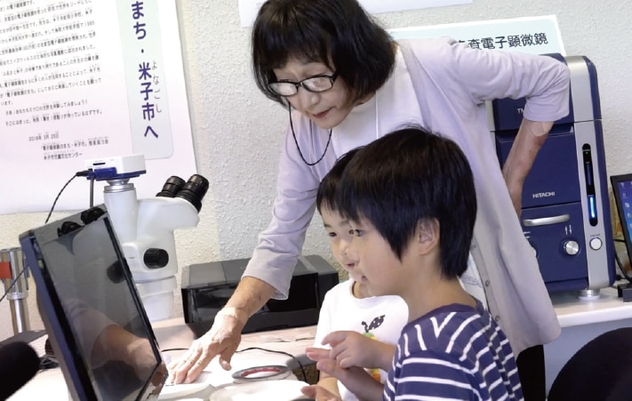
Doctor Inaga guiding students through an observation. Volunteers who have taken a training course offered by members of the project committee may serve as guides in rotation to help children make observations.
INAGA: That’s right! Kaname Ishikura was in his second year of middle school when we opened the facility, and he wound up spending so much time there that now he can operate the microscope by himself. He’s also a gifted writer who has entered many writing competitions, and just the other day we learned that his essay “Inheriting the challenge of the microscopic world,” which he submitted to the contest for Sankei Shimbun’s 2020 Culture Award for high-school students, had won an Excellence Award—the second-highest prize in the national competition. His essay described his experience, starting in middle school, of using electron microscopes to observe specimens; he also discussed his interactions with Professor Tanaka and with me, and expressed a firm commitment to following in Professor Tanaka’s footsteps as an explorer of the microscopic world. If our decision to install an electron microscope at the Children’s Center played any role in stoking Kaname’s interest in science, that would be a truly wonderful reward.
Meanwhile, Chiaki Muku, a primary-school student, captured an image of secretory gland structures on the surface of a mini tomato that won an “Honorable Mention” in the Japanese Society of Microscopy’s 2019 photo contest. The image very clearly depicted protrusions closely resembling four leaf clovers, and was recognized by an Honorable Mention even though the contest usually selects only one winning image. The news that a second-grader had pulled off such an impressive accomplishment was a hot topic of conversation throughout the region.
In capturing microscopy images, children are often able to succeed where we researchers fail due to our preconceived notions and biased thinking; when this happens, we find it tremendously stimulating. Professor Tanaka always said that he wanted children to appreciate the beauty of the microscopic world—and to experience it for themselves; as we move toward turning this dream into reality, I like to think he’s up there somewhere looking on with pride and joy.
KOGA: To me it seems clear that this project could only have been successful with Professor Inaga at the helm. My own efforts to expand awareness of electron microscopy—which seem almost embarrassingly feeble by comparison—have focused on mass-media strategies; for example, with help from the folks at Hitachi High-Tech I recently appeared on an NHK science program. I should say that, in general, presenting to an audience is one of my least favorite things to do, and I’m not even sure that performing on TV or in other media is the right strategy, so in the past when I was asked to provide photographs or appear in public I always declined. However, eventually someone encouraged me to make more of an effort—“as part of an outreach effort to promote awareness of electron microscopy among the general public,” they said—and I began to reevaluate my reticence.
One current societal trend that I find concerning is “detachment from electron microscopy.” I think an effective way to counteract this trend is to reach out to children—through the media, of course—to get them interested and engaged in the microscopic world. Of course this isn’t something I can do on my own—we need help from the good folks at Hitachi High-Tech and many other people—but I’ve become committed to outreach initiatives to convince even just one more person of the enormous appeal of our field.
INAGA: In contrast to our work promoting Yonago: the electron-microscope town—whose impact is obviously limited to a single geographical region, Professor Koga’s work has the potential to reach people all throughout Japan—and around the world. I think both types of outreach are important. People find Professor Koga’s microscopy images to be convincing precisely because they are so spectacular—it’s impossible to look at them without being moved by their beauty. Going forward, I’ll keep doing everything I can to sound the message far and wide: Beauty is truth, truth beauty.
KOGA: Thank you very much for those kind words.
INAGA: That’s correct. The work we do to promote Yonago: the electron-microscope town has sometimes involved working with artists, and in March 2019, to celebrate the one-year anniversary of our founding, we presented exhibits at the Children’s Center and at the Yonago City Museum of Art displaying a collection of over 100 electron microscopy images captured by children. One of the visitors to our exhibition was the artist and musician Tomoya Matsuura, whose work involves SEM images presented as works of art—and who, we heard, had submitted an entry to the Japanese Society of Microscopy’s photo contest. By coincidence, this happened to be the year in which an image of HeLa cell chromosomes submitted by me and my collaborators at Hitachi High-Tech won first prize in the contest, so it was through that connection we wound up working with Mr.Matsuura.
Later that year, at a conference on chromosomes in September 2019, Mr. Matsuura and I presented an artistic creation consisting of a movie made from our HeLa cell micrographs and set to music; this communicated research results, not through papers, but through sound and images, and was a big hit with the audience. Right now we’re collaborating with Mr. Matsuura to create video content in which electron-microscopy images are paired with music; we plan to show the completed product in the planetarium at Yonago Children’s Center.
Mr. Matsuura’s work exudes an artistic beauty that captivates many viewers—including, for example, the organizers of the BIWAKO BIENNALE 2020 art festival, who used a SEM image captured by Mr.Matsuura as the background of the festival’s official poster. Looking at his creations, one has the sense of being present at the creation of a promising new domain of applications for electron microscopes—as tools for artistic expression.
KOGA: I agree that there’s a sense in which SEM images can be interpreted as artworks. Black-andwhite images give a feel for the structural beauty of nature, while adding color takes you to totally different worlds. These days we add color to enhance the visual clarity of images for the sake of academic accuracy, but with more time I’d love to explore more artistic coloration schemes.
INAGA: For me, as I said before, the main goal is to organize all the research I’ve done thus far. As a matter of fact, I used some of my retirement pay to purchase a Miniscope® for myself, and even after my position as a project researcher comes to an end I plan to continue doing research just like Professor Tanaka did. However, working with chromosomes really requires a FIB-SEM system, so for the time being my immediate goal is to organize all the data I’ve gathered thus far. I’d also like to work on establishing low-vacuum SEM techniques for renal biopsy analysis that would be easy to implement at clinics, to help make this approach more common.
I’m also hoping to continue moving forward with our work promoting Yonago: the electron microscope town. Recently we’ve been excited about the idea of running an electron-microscopy image contest, open to pros and amateurs alike, to celebrate our three-year anniversary.
KOGA: There are a number of things I’d like to do going forward, but the one most closely related to our discussion today is to establish osmium maceration as a method that anybody can use. When things go well, osmium maceration really does allow you to capture beautiful images. However, although many people understand this, a lot of times they say it’s just too hard and give up—and some even grumble that Koga’s the only one who can get it to work. But the reality is that, even after working on it for more than 10 years, I’ve only recently started to feel like I’m getting the hang of it.
The basic idea of osmium maceration (as we discussed in Part One) is to use a thin osmium liquid to remove soluble proteins while selectively retaining cell membranes. After a lot of my own trial-and-error experiments, I’ve come to understand that the temperature settings for the maceration procedure are crucial. Professor Tanaka recommended keeping the temperature fixed at 20°C, but I’ve found that slightly higher temperatures allow the maceration time to be reduced with almost no failures, and I was able to present this in a research paper. Going forward, I’d like to revisit the fixing fluid conditions to simplify things and make the method into something that anybody can recreate with good stability.
If my work on this helps make osmium maceration a more common technique, I’ll feel like I have repaid some of my debt to Professor Tanaka. I dream of visiting the Professor’s grave to let him know that osmium maceration has become a technique that anybody can use.
INAGA: Professor Koga, a while back you and I went together to visit Professor Tanaka in the hospital, and I remember that he talked to us for an unusually long time and was full of praise and encouragement for our work. And yet, toward the end, he said “But still it seems like there might be something missing...” Do you remember that?
KOGA: Yes, I remember that well.
INAGA: Whenever I would report some new research achievement, I remember Professor Tanaka would always congratulate me—but then would always repeat that same sentence at the end. I think he was trying to say “Never be satisfied with any single success, and always keep striving toward your goal.” I’m pretty sure that this is what he said to himself, and that it’s what motivated him to work so hard for so long.
So, whenever I’m feeling happy about something that has gone well, I try to remember the Professor’s advice—and, Professor Koga, I suspect you’ll always continue to hear Professor Tanaka saying the same thing to you. Never be satisfied with the status quo, and always keep searching for the next round of answers. This disposition as a scientist is part of Professor Tanaka’s legacy, and I think we have a duty to pass it on to future generations whenever and wherever we can.
(Interview and text: Akiko Seki)
Original paper written in Japanese "Continuing the Mission of Professor Keiichi Tanaka: Pursuing—and Communicating—Beauty in the Form of Truth (Part Two)" has been translated to this English version by Hitachi-High Tech Corporation.