
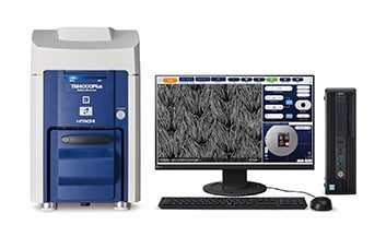
Tabletop Microscopes TM4000PlusIII/TM4000III
A tabletop microscope for the next generation of scientific leaders is now available
The TM4000PlusIII/TM4000III is the newest addition to a lineup of microscopes that have sold over 5,800 units worldwide.
Our newest updates bring improvements to users at the forefront of R&D, quality control, and education.
Features of TM4000PlusIII/TM4000III
Reduce Your Workload and Achieve more consistent results
Automated observation of multiple specimens: Recipe Control*1
The TM4000PlusIII recipe function can be used to automatically execute operations, such as stage movement, magnification setting, and image capturing. Once the user builds a recipe, it can be executed to run the tool automatically with no further intervention. This significantly reduces the workload on operators and allows for more consistent results, even from inexperienced users.
Faster automatic particle analysis: High-current function + AZtecLiveLite particle analysis package*
Tasks such as purity analysis of industrial products and particle analysis of filters can be time consuming as data is required from many locations to complete the analysis. The TM4000PlusIII is equipped with a new high-current setting, Mode 5, which provides increased signal for imaging and EDS analysis. In combination with the Oxford Instruments EDS AZtecLiveLite particle analysis package, this helps to increase throughput.
Field-of-view navigation with less noise: High-current function
SEM field-of-view navigation is performed using fast scanning modes, so images tend to be prone to noise. The high-current function allows for easy field-of-view navigation due to its high signal-to-noise (S/N) ratio. This takes the stress out of specimen navigation!
Plan Ahead with the Filament Monitoring Function
Maintenance planning: Filament monitoring function
The TM4000PlusIII is equipped with a system that monitors the filament and displays graphics to show the remaining lifetime of the filament. If the equipment is being operated by multiple users, this makes it possible to plan the use of the tool in a systematic way. There is no more need to worry about the filament suddenly running out when the tool is most needed.
Ideal for Educational Purposes
Experience the world of electron microscopy with no preprocessing required:
Low-vacuum function + high-sensitivity backscattered-electron detector
Users can try out electron microscopy with all kinds of specimens: picked flowers, treasured minerals or familiar foods, with no need for metal coating. The low-vacuum function of the TM4000PlusIII/TM4000III reduces preprocessing, and the high-sensitivity backscattered-electron detector (BSED) allows images to be obtained quickly.
Build programming skills with workflow automation functions (TM4000PlusIII)*1
Developing and maintaining digital skills is vital in today’s world. The automation support function allows you to experientially learn important programming concepts such as "sequential execution," "repetition," and "conditional branching" through the operation of the TM4000PlusIII.
*1 Optional for TM4000PlusII only
*2 Optional
Features of Tabletop Microscope
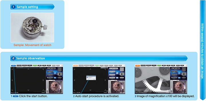
A quality image can be obtained with simple steps.
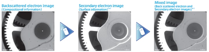
Automation, Observation, and Elemental Analysis
Easy to switch images with one-click.
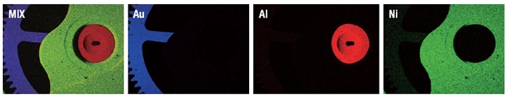
Rapid acquisition of elemental maps *2
Sample: Movement of watch
*1 Secondary electron images and MIX images can only be observed in TM4000Plus III
*2 Option
Intuitive operation on Camera Navi
Use of optical images helps navigate to target observation area easily.
Obtained SEM images can be layered on a SEM MAP image.
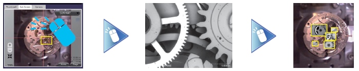
Sample: Movement of watch
Camera Navigation System
Report Creator
Simply select images and a template to create customized reports.
Created reports can be saved/edited in Microsoft Office® formats.
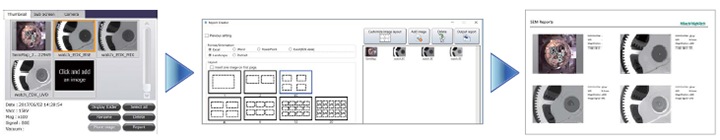
Sample: Movement of watch
The image on the screen includes options.
Imaging non-conductive samples without sample prep
Charge-up reduction mode
Charge on a sample can be reduced by one-click.
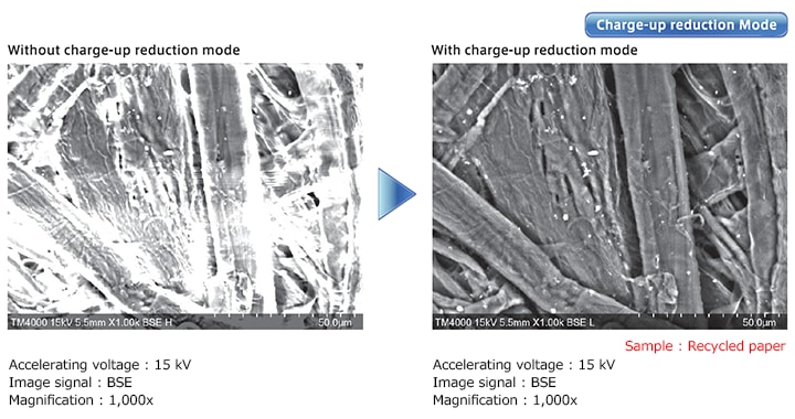
Image a variety of materials under low vacuum condition
The images show observations of non-conductive samples such as ink toner particles and a hydrated leaf surface.
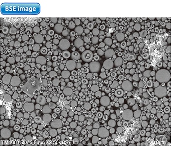
Accelerating voltage: 5 kV
Image signal: BSE
Magnification: 2,500x

Accelerating voltage: 10 kV
Image signal: SE
Magnification: 100x
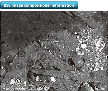
Obtaining SE images in low vacuum mode
Innovative secondary-election detector to obtain surface detail with non-conductive samples at lower vacuum conditions
The TM4000PlusIII can observe not only conductive samples, but also non-conductive or hydrated samples without sample preparation. Switching between BSE and SE can be performed easily.
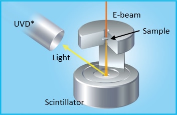
High-sensitivity Low-vacuum SE Detector (UVD)
Hitachi's UVD generates secondary-electron images by detecting visible light excited by electron gas interactions.
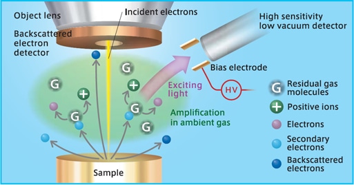
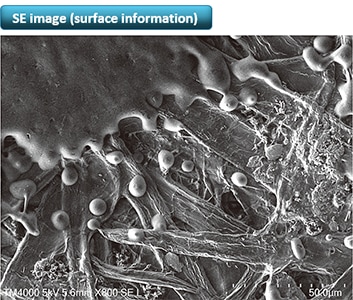
Image signal: SE
Magnification: 800x
Accelerating voltage: 5 kV
Image signal: BSE
Magnification: 800x
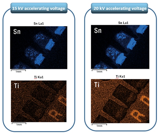
Advantages of 20 kV accelerating voltage
High accelerating voltage enables higher-speed EDS analysis.
EDS mapping data at 20 kV in 2 min
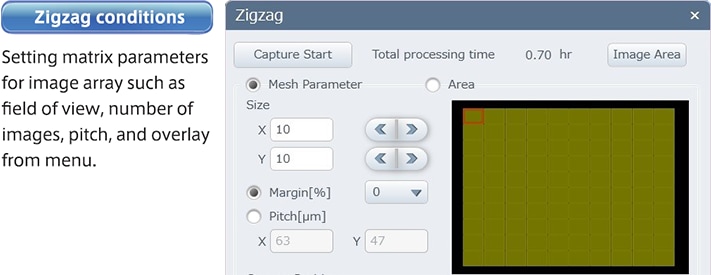
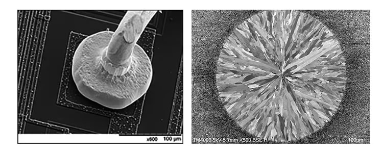
Multi Zigzag (Option)
A function that takes multiple high-magnification images and stitches them together to create a single high-resolution image.
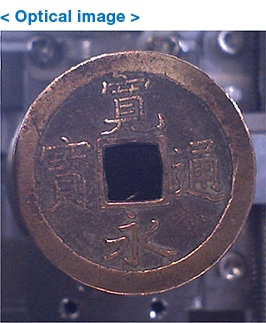
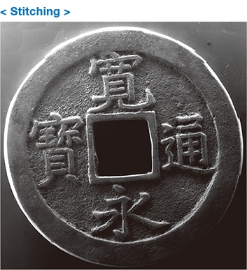
Accelerating voltage: 15 kV
Image signal: SE
Magnification: 30x
Field of view 10 vertically × 12
horizontally
(some parts were trimmed)
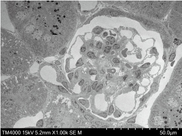
STEM holder (Option)
Easily obtain transmitted images on thin samples
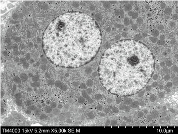
The newly developed STEM holder can be used to perform transmission images with the Hitachi UVD. Images of thin or biological samples can be obtained.
* UVD is a function of TM4000III.
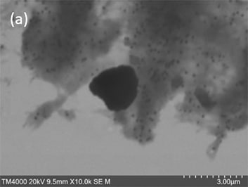
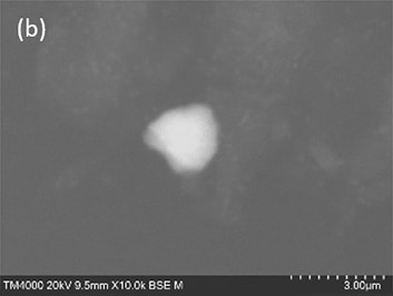
Accelerating voltage: 20 kV
Image signal: (a) STEM, (b) BSE
Magnification: 10,000 x
Accelerating voltage: 15 kV
Image signal: STEM
Magnification: 1,000 x
Accelerating voltage: 15 kV
Image signal: STEM
Magnification: 5,000 x
Applications Gallery
Specifications
| 项目 | TM4000PlusⅢ | TM4000Ⅲ |
|---|---|---|
| 倍率 | ×10~×100,000(照片倍率) ×25~×250,000(显示器倍率) |
|
| 加速电压 | 5 kV、10 kV、15 kV、20 kV | |
| 图像信号 | 背散射电子 二次电子 复合图像(背散射电子+二次电子) |
背散射电子 |
| 真空模式 | 导电体(仅背散射电子) 标准 荷电减轻 |
标准 荷电减轻 |
| 样品可移动范围 | X:40 mm Y:35 mm | |
| 最大样品尺寸: | 80 mm(直径) 50 mm(厚度) | |
| 电子枪 | 预对中钨灯丝电子枪 | |
| 检测器 | 背散射电子:高感度4分割 背散电子检测器 二次电子:高感度低真空 二次电子检测器 |
背散射电子:高感度4分割 背散射电子检测器 |
| 排气系统 (真空泵) |
涡轮分子泵:67 L/s 1台 隔膜泵:20 L/min |
|
| 马达台 | 自动马达台 | 手动样品台 |
| 尺寸与重量 | 主机:330(宽)×614(纵深)×547(高)mm 55 kg | 330(宽)×617(纵深)×547(高)mm 53 kg |
| 隔膜泵:144(宽度)×270(纵深)×216(高度)mm | ||
 Powered by Bioz